Research
“What I cannot create, I do not understand” - Richard Feynman
Our lab works on this basis coming from a reductionist approach, piecing together minimal components from a cell to recreate the patterns seen inside the cell.

Bacterial Cell Division
It has been recently discovered that bacteria have cytoskeleton and exhibit highly dynamic patterns. We are interested in understanding how bacteria, despite their small size are able to spatially organize genomic DNA and proteins to specific locations in the cell. These bacterial positioning systems form distinctive patterns within the cells, and are involved in important cellular functions such as DNA segregation, cell division and bacterial motility. We use multidisciplinary techniques such as single-molecule imaging, synthetic biology and microfluidics to study the molecular mechanisms of spatial organization using cell-free systems.

Chromosome dynamics
Chromosome replication and segregation are essential processes in the cell cycle. To ensure that each daughter cell receives a copy of the genome, sister chromosomes must be accurately distributed in space and time to opposite cell halves. We are interested to understand the molecular mechanisms that are involved in the physical movement of DNA and macromolecular complexes in the cell.

Cell-free Reconstitution
In vitro reconstitution is a powerful techniuqe used to study the fundamental mechanisms that drive the cellular processes of a biological system. We use a reductionist approach to recreate a minimal biological system from purified components and to identify the conditions required to reproduce the cellular dynamics.

Single-molecule fluorescence microscopy
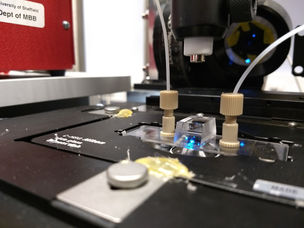
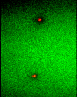
We have built a dual-color TIRF microscope that is capable of imaging protein-protein/DNA interactions at the single-molecule to mesoscale level. It is connected to two syringe pumps that allows the infusion and mixing of samples into microfluidic flow cells that we can directly observe on the microscope.

Tau protein aggregation
Tau is an essential protein in the brain and plays a key role in the pathology of a range of neurodegenerative diseases, including Alzheimer’s. Tau is an intrinsically disordered protein that binds microtubules to stabilize the cytoskeleton of nerve cells. We are interested in studying the aggregation of Tau and its cellular interactions. How does Tau protein self-assemble to form abnormal aggregates and filaments that is the pathological basis of Alzheimer's? We use single-molecule imaging and a range of biochemical techniques to investigate Tau dynamics and interactions.


